What Are The Colors On Thyroid Ultrasound
Colour is blue or red depending on whether the blood movement is towards the ultrasound probe or away from it. On color doppler, the inferior thyroid artery (arrow) is seen, (c) blood flow pattern in normal thyroid gland.
Doppler Ultrasound Radiology Key

The thyroid gland is located in front of the neck just above the collar bones and is shaped like a butterfly, with one lobe on either side of the neck connected by a.

What are the colors on thyroid ultrasound. Grayscale and color doppler ultrasound imaging of the thyroid gland. Doctors apply a gel, which allows the sound waves from the ultrasound probe to get through the skin. It is not uncommon for one lobe to be larger then other;
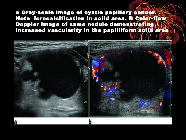
An ultrasound of the thyroid produces pictures of the thyroid gland and the adjacent structures in the neck. Ultrasonography can deliver a diagnostic accuracy of over 90% in thyroid carcinoma, especially papillary carcinoma. Then go to the right neck and take sagittal images in middle of the right thyroid lobe, lateral.
Communicate with your technician if you experience any discomfort. (2000) color flow doppler sonography of the thyroid. If a biopsy is needed, doctors use ultrasound to scan the area again, ensuring that they know exactly where to place the needle.
Also place color doppler over the gland. This nodule (shown in red) comprises about 80% of the thyroid tissue (shown in yellow) in this particular area of the thyroid. The red and blue indicates movement of blood through vessels.
A thyroid scan is a simple outpatient procedure. The probe detects these reflections to make pictures. Fnac was advised and would show a classic gritty feel as.
During thyroid us, transverse and longitudinal images should be obtained, while color doppler images may be useful in selective cases (6). Thyroid ultrasounds with rule out parathyroid disease: The hashimoto thyroid as well as subacute thyroiditis will cause heterogeneous echotexture [your ultrasound report says homogeneous], and the lobe itself will be tender, not soft.
Transmit ultrasound, it must be coupled to the skin with a liquid medium or a gel. Dark spots could represent thyroid nodules or growths. Bogazzi f., bartalena l., martino e.
Have the patient look to the left side when scanning the right neck. In the more frequently used color map, the color red represents hard or stiff tissue and the color purple represents soft or elastic tissue. Thyroid hypoechogenicity at ultrasound is a characteristic of autoimmune thyroid diseases, with an overlap of this echographic pattern in patients affected by graves' disease or hashimoto's thyroiditis.
In addition you will have swelling on the right side only. (a) gray scale ultrasound, transverse scan showing normal thyroid anatomy, (b) arterial vascularization of the thyroid gland. You lie flat on the table, with your head and neck extended.
In our experience, most benign thyroid nodules are either purple or green. Take image of the thyroid at midline, sweep superior and inferior and measure the isthmus in anteroposterior dimension. Color doppler ultrasound shows typical twinkling artefacts in this calcific colloid nodule of the thyroid.
Images will be visible on a screen, and are used to make sure that the radiologist has a clear picture of your thyroid to. On spectral display, a low resistance flow with a high peak systolic velocity is obtained The color green indicates the median stiffness.
If you looked at other parts of the thyroid, however, you would not. The signal is organized electronically into numerous shades of gray It is generally normal unless there is too much color, which would have been mentioned in the report.
Document any parathyroid glands if seen and measure in 3 dimensions. Blue represents that the blood flow is away from the probe and red represents that the blood flow is towards the probe. Vascularity noted on color doppler imaging.
Publisher name springer, boston, ma; You need to talk to the doctor who ordered the ultrasound so you will know for sure what the u. Color on your thyroid ultrasound means that color doppler was applied and blood flow was detected.
(example abnormal calcium levels) (please document the thyroid as well using the thyroid protocol) images: This instrument rapidly alternates as the generator of the ultrasound and the receiver of the signal that has been reflected by internal tissues.
![]()
Hashimotos Thyroiditis Transverse Gray-scale Ultrasound A And Color Download Scientific Diagram

Graves Disease Radiology Reference Article Radiopaediaorg

Thyroid Doppler Ultrasound During Thyroid Storm On Grepmed
Pdf Gray-scale Vs Color Doppler Ultrasound In Cold Thyroid Nodules Semantic Scholar
Pin On Sonography

Pin On Thyroid Gland
Normal Ultrasound Of Thyroid Gland And Lymph Nodes Radiology Key


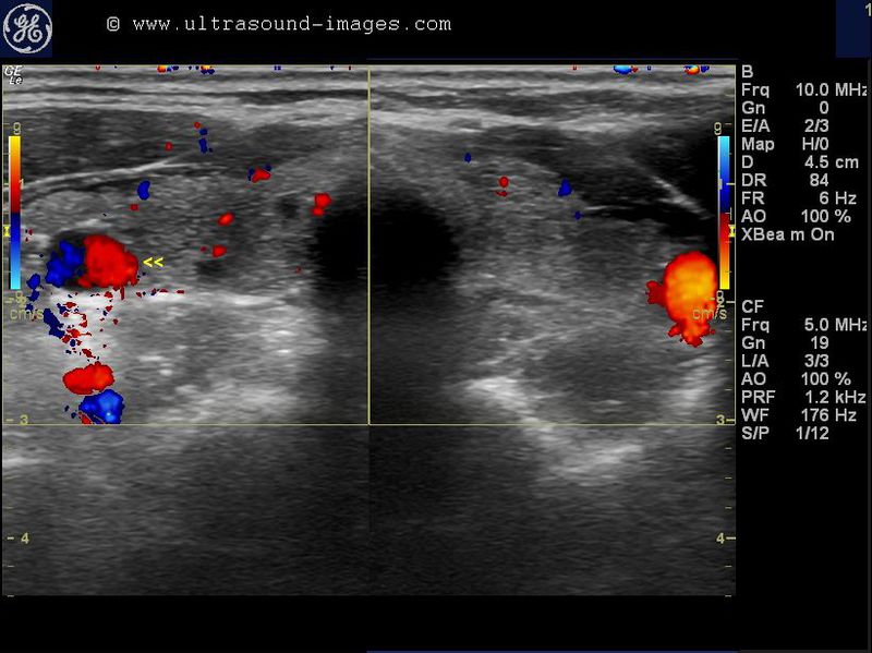
A And B Case 3 Ultrasound With Color Doppler A Left Lobe Thyroid Download Scientific Diagram

Conventional And Color Doppler Thyroid Ultrasound Effectiveness And Evaluation Thyroid Ultrasound Mohammed Ahmed M Awad Alkareem Dr Rania 9781726756112 Amazoncom Books

Hashimotos Thyroiditis And A Giraffe Pattern On Thyroid Ultrasound
A Gallery Of High-resolution Ultrasound Color Doppler 3d Images - Thyroid-2
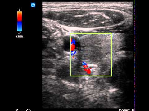
Transverse Color Doppler Sonogram Of A Nodule In The Right Thyroid Lobe Download Scientific Diagram

Thyroid Nodule Ultrasound What Is It What Does It Tell Me - Quick Painless Inexpensive And Accurate
Thyroid Ultrasound

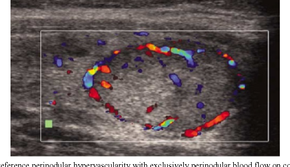
Intranodular Vascularity Grading On Color Doppler And Representative Download Scientific Diagram

Color Doppler Patterns A Pattern 0 Normal Thyroid Vascularity B Download Scientific Diagram

Ultrasound Of The Thyroid Showing An Intense Vascularity On Color Download Scientific Diagram

Pin On Tirads Us Thyroid
Normal Thyroid Color Doppler - Youtube
